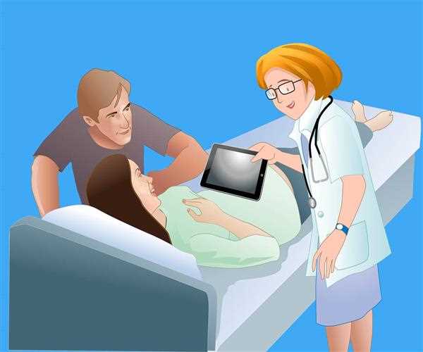
Your doctor or qualified technician will use a plastic transducer to transmit high-frequency sound waves through your ****** during pregnancy. These sound waves are fed back into the machine, where they are converted into pictures of your baby.
Ultrasound can help your doctor monitor your baby's progress, diagnose abnormalities, estimate your due date, determine if you are carrying multiple items, and reveal the condition of your placenta. You can set and set your child's ****, among other things.
To help you prepare for these important tests, we're breaking down the most common prenatal ultrasounds weekly, as well as when you should be guessing when.
Ultrasound in the first trimester of pregnancy (6-8 weeks)
When you are six to eight weeks pregnant, you may get your first ultrasound, often referred to as a baby sonogram. However, not every woman has this scan; Some doctors can only do this if the woman has specific high-risk pregnancy symptoms such as bleeding, abdominal discomfort or birth abnormalities, or miscarriage.
An early pregnancy ultrasound can be done transvaginally to allow professionals to see your baby more clearly. In this situation, your OB-GYN inserts a thin wand-like transducer probe into your vaginal canal, sending high-frequency sound waves into your ******. Sound waves resonate with the fetus, sending information to a computer that converts the images into a black-and-white image of your baby.
The baby's heartbeat can be seen during the first six weeks of pregnancy. Your doctor will also estimate your baby's due date, track milestones, count the number of babies in your **** and check for an ectopic pregnancy.
Ultrasound Dating (10-13 weeks)
Those who skip the ultrasound in six to eight weeks can get a 'dating ultrasound' between 10 and 13 weeks. The expiration date, your baby's 'crown - **** length' (measured from head to bottom), the number of babies in *, and the fetal heart rate are all provided.
Ultrasound of Nuclear Translucency (14-20 weeks)
According to John Stone, a professor of obstetrics, gynecology, and reproductive science at the Mount Sinai School of Medicine in New York, you should have a nocturnal translucency (NT) test between 14 and 20 weeks for Down syndrome and heart problems. Could not. , Or other chromosome abnormalities. If your screening test detects a potential problem, you can take it if you are 35 or older or have a family history of specific birth abnormalities.
During nocturnal translucency screening, the doctor uses an ultrasound to determine the thickness behind the baby's neck (a blood test is also used to detect hormones and proteins). A large neck indicates a higher probability of congenital abnormalities such as Down syndrome.
Anatomy scan (18-20 weeks)
This comprehensive prenatal ultrasound, usually done between 18 and 20 weeks of the second trimester, lasts 20 to 45 minutes for the baby and lasts for several days. This is the most complete test you can get before your baby is born.
According to Zen Chooh, director of prenatal diagnosis and medicine at Lucille Children's Hospital Stanford in Palo Alto, California, doctors check your baby's heart rate and look for abnormalities in their brain, heart, kidneys, and liver during an anatomy scan. , Your baby's fingers and toes will be counted, birth abnormalities will be checked, the placenta will be examined and amniotic fluid level will be measured. They can find out exactly the gender of your baby, though it is not given.
Ultrasound in the third trimester
In the third trimester, most parents do not need an ultrasound. According to Dr. Chooh, if your pregnancy is considered high-risk - for example, if you have high blood pressure, bleeding, low levels of amniotic fluid, premature contractions, or more than 35 - your doctor should consult your doctor. In the office during your previous visits for assurance, ultrasounds can be done with a lower resolution. If your 20-week scan is covered with your ****** Maya, you should undergo a follow-up ultrasound.
Other ultrasounds for pregnancy
Your doctor may perform other pregnancy tests that may require an ultrasound. Two examples are chorionic villus sampling (CVS) and amniocentesis. Ultrasound is also used in fetal echocardiogram, which displays the baby's heartbeat and confirms complications.


Leave a Comment