Fluorescent sensors, which are most probably used to label and image a wide variety of molecules actually offers a unique glimpse of living cells. However, these can only be used within cells which have matured either in a lab dish or in tissues which are close to the surface of the body. It is such because their signal gets lost once they are implanted too deeply.
HIGHLIGHTS
- Engineers found a way to improve the signal emitted by fluorescing nano-sensors.
- The researcher’s technique allows particles to be placed deeper within the tissue.
- The frequency of fluorescent light which is emitted by the sensor can be modulated.
Well, MIT engineers are noticed to come up with a way through which they can overcome such limitation. A novel photonic technique which they have developed is used for exciting any fluorescent sensor which could dramatically improve the fluorescent signal. With this approach, the researchers have showcased that they could implant sensors as deep as around 5.5 centimeters in tissue and still get a powerful signal.
The researchers have mentioned that such technology may enable fluorescent sensors that can be used to track specific molecules which are inside the brain or some other tissues as well which are deep inside the body especially for diagnosis or observing drug effects.
Volodymyr Koman, an MIIT research scientist and one of the lead authors of the new study have said “If you got a fluorescent sensor which will probe biochemical information in cell culture, or in thin tissue layers, this technology permits to translate all of the fluorescent dyes and probes into thick tissue.”
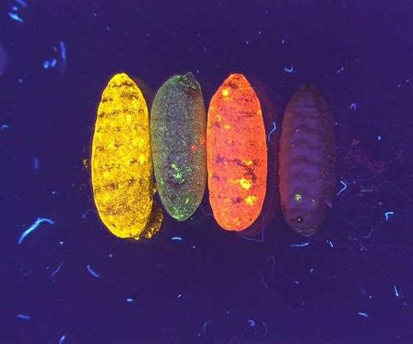
Scientists use many alternative forms of fluorescent sensors like quantum dots, carbon nanotubes, and fluorescent proteins, to label molecules within the cells. These sensors’ fluorescence is noticed by shining laser light on them. However, this light does not work in thick, dense tissue, or deep inside the tissue. It is because the tissue itself emits some fluorescent light. This light is therefore, known as fluorescence, drowns out the signal which is coming from the sensor.
“All tissues autofluoresce later becomes a limiting factor,” Koman says. “As the sensor’s signal get weaker with time, it becomes overtaken by the tissue fluorescence,” he added.
In order to overcome this limitation, the MIIT team has come up with a way to modulate the frequency of the fluorescent light which is emitted by the sensor so it is distinguished from the tissue autofluorescence. This technique is named as wavelength-induced frequency filtering (WIFF), which uses three lasers to form a laser beam with an oscillating wavelength.
Also Read: Scientists Say 'Alien' Rock Hypatia May Reveal Details Leads to Supernova Explosions